Home > Science > SEM
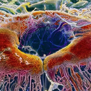
Eye anatomy, SEM

![]()
Wall Art and Photo Gifts from Science Photo Library
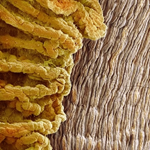
Eye anatomy, SEM
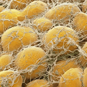
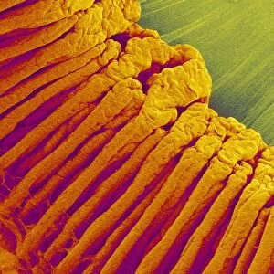
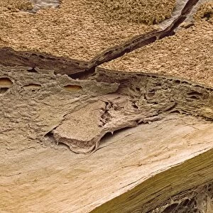
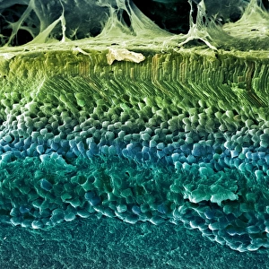
Eye anatomy. Coloured scanning electron micrograph (SEM) of part of the ciliary body (red/yellow) of the eye, a ring-shaped structure inside the eye, surrounding the iris. This view looks at part of the ciliary body (arching from top to bottom down right), as seen from inside the eye. Part of the iris is seen down left. Part of the choroid (outer layer of the eye) is just seen at far right. The ciliary body joins to ligaments that hold the lens in place behind the iris. The lens has been removed here. The ciliary body also contains the ciliary muscle that is contracted to alter the curvature of the lens and focus light on the retina at the back of the eye. These structures are at the front of the eye
Science Photo Library features Science and Medical images including photos and illustrations
Media ID 6449465
© SUSUMU NISHINAGA/SCIENCE PHOTO LIBRARY
Choroid Ciliary Body Inside Internal Iris Ligaments Muscles Ocular Ophtalmological Ophthalmology Physiological Physiology Tissue False Coloured
FEATURES IN THESE COLLECTIONS
MADE IN THE USA
Safe Shipping with 30 Day Money Back Guarantee
FREE PERSONALISATION*
We are proud to offer a range of customisation features including Personalised Captions, Color Filters and Picture Zoom Tools
SECURE PAYMENTS
We happily accept a wide range of payment options so you can pay for the things you need in the way that is most convenient for you
* Options may vary by product and licensing agreement. Zoomed Pictures can be adjusted in the Cart.